
Persistent Ocular Symptoms After COVID-19 Point to Neuroinflammation
07/09/2026
Key Takeaways
- Photophobia, eye pain, eye fatigue, and focusing difficulty were commonly reported, and symptoms often persisted from months into several years.
- Routine anterior-segment and refraction measures were largely unchanged, while near-vision, corneal sensory and nerve, pupillary, and mature dendritic/T-cell findings differed on specialized testing.
- Tear proteomic dysregulation and cohort-derived classification models were observed, and the authors interpreted the overall pattern as consistent with neuroinflammatory, autonomic, and peripheral ocular nerve involvement.
This prospective cross-sectional study used single-visit ophthalmic assessments at Linköping University Hospital between April 2022 and September 2023. Photophobia, eye pain, eye fatigue, and reduced focusing ability were among the most common complaints, and symptom onset occurred 0.2 to 4 months after infection in 68%. Time from first infection to examination ranged from 3 to 42 months, 33.3% had symptoms for at least 2 years, 73% were female, and six participants were younger than 18. Modified Catquest-9SF scores showed worse vision disability in POS than controls, at −0.65 ± 2.26 versus −5.44 ± 2.00 logits, with P<0.0001. One-third were on part-time or full-time sick leave, and only 39% carried a formal long-COVID diagnosis.
Routine anterior-segment and refraction testing did not differ across several standard measures, but near-vision testing separated the groups. Best-corrected binocular near visual acuity differed by +0.038 logMAR in POS, with P=0.0047. Near ocular alignment and fixation differed by +1.29D, with P=0.0008, and near vision fusional reserve was lower by −5.38D, with P=0.014. Uncorrected monocular distance visual acuity also differed by +0.19 logMAR, with P=0.022.
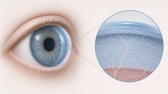
Specialized measures identified differences in corneal nerve, sensory, autonomic, and immune-related domains. Corneal subbasal nerve density was lower by −2.85 mm/mm2, with P=0.0001, and central corneal esthesiometry showed a weaker blink reflex, requiring 1.79 mm shorter filament length, with P=0.0022. Dynamic pupillometry showed larger dilated pupil size at +0.47 mm, with P=0.026, and longer constriction time at +0.09 s, with P=0.013. Mature dendritic/T-cell density was higher by +8.05 cells/mm2, with P<0.0001, whereas immature cell density did not differ significantly.
In the tear proteomic analysis, investigators identified 178 dysregulated proteins, and five remained significant after FDR adjustment: ITGB6, NFASC, CKMT1A-CKMT1B, CTGF/CCN2, and TPSAB1. A five-parameter clinical model reached a cross-validated AUC of 0.77, which rose to 0.91 after six tear proteins were added in cohort-derived penalized logistic regression. Objective diagnostic criteria for POS are not established, and tear-film diagnostics are not standard testing, so both models remain bounded by those conditions.